Pharmacology of the Autonomic Nervous System (ANS)
The ANS is part of the Peripheral Nervous System (PNS).
The PNS consists of 3 principal elements:
1. Autonomic nervous system
2. Somatic efferent system, innervating skeletal muscle
3. Somatic and visceral afferent system
Arrangement of neurons
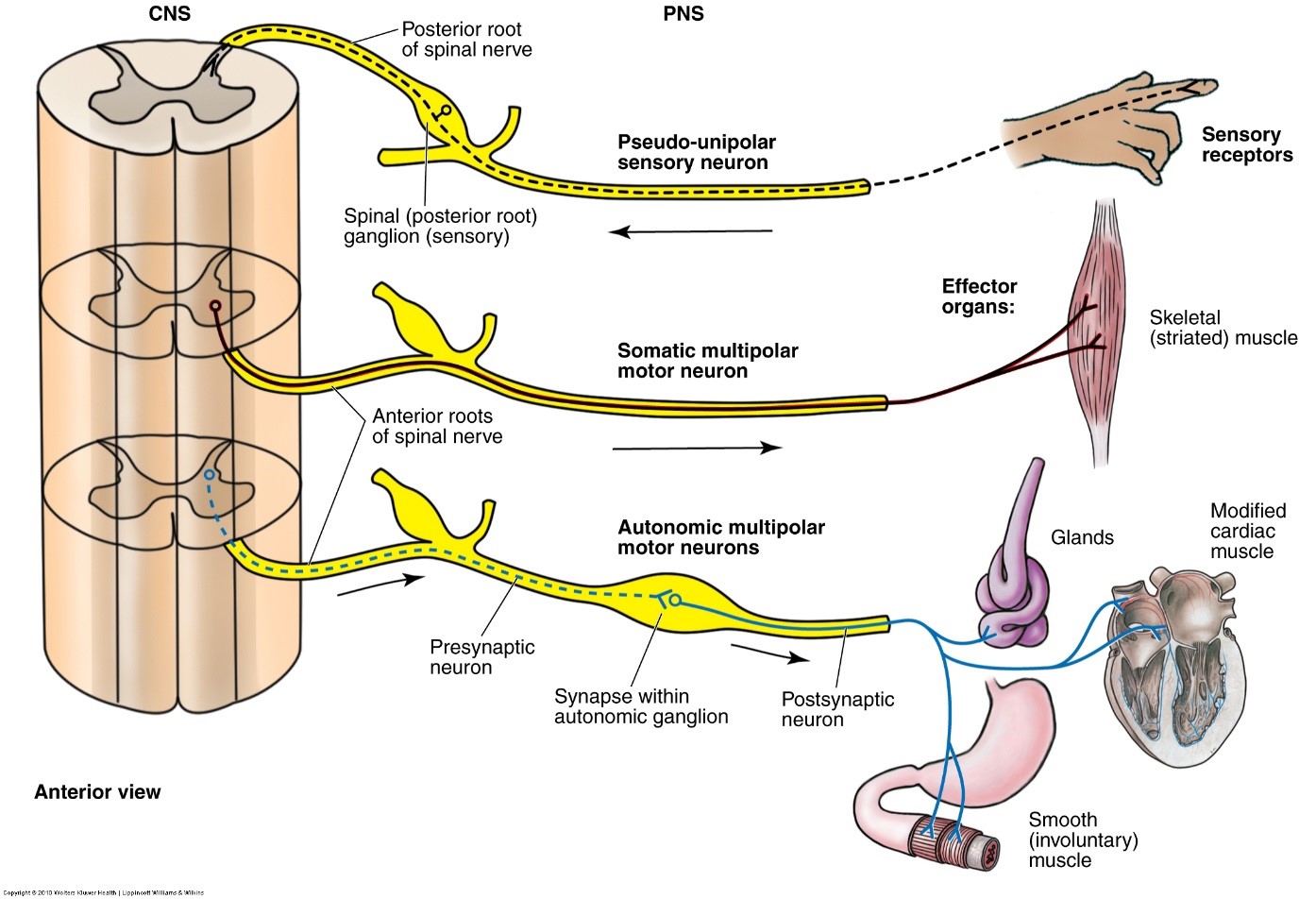
- In the somatic efferent system a single motor neurone connects the CNS to the muscle fibre.
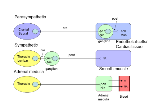
- In the autonomic pathway there are two neurones, preganglionic and post-ganglionic, which synapse in an autonomic ganglion outside the CNS.
Video by Dr James Pickering -
https://www.youtube.com/watch?v=WXO_nyFY9oM&index=16&list=PLrSF4J33FBEjtWaLRVW6DNy6BKCOatf0A
Organisation of the ANS
The ANS is largely outside the influence of voluntary control
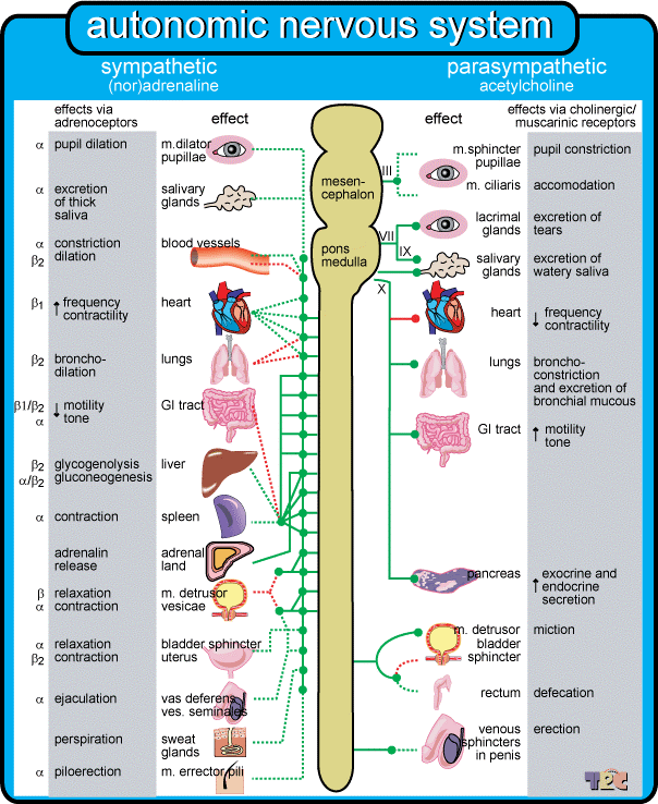
The ANS regulates:
- Contraction and relaxation of smooth muscle
- All exocrine and some endocrine secretions
- The heartbeat
- Certain steps in intermediary metabolism
Potential pharmacological targets
- Inhibition of transmitter synthesis
- Inhibition of transmitter release
- Promotion of transmitter release
- Inhibition of transmitter storage
- Inhibition of transmitter re-uptake
- Inhibition of transmitter metabolism
- Inhibition of postjunctional ganglionic receptors
- Inhibition of postjunctional effector receptors
Cholinergic receptors
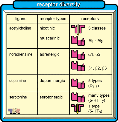
- Cholinergic receptors respond to the neurotransmitter acetylcholine (ACh)
- 2 types of ACh receptor were originally classified on the basis of sensitivity to the alkaloids muscarine and nicotine that mimicked some, but not all, of the actions of ACh.
Nicotinic receptors
- Nicotinic receptors are located postsynaptically in all autonomic ganglia and at the neuromuscular junction (covered in muscle and nerve lectures)
- At these junctions nicotinic receptors function as the excitatory receptor for the postsynaptic cell.
- Release of a sufficient quantity of ACh from the adjoining presynaptic cell causes an excitatory response in autonomic ganglion cells and in somatic muscle fibres.
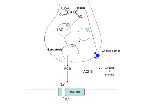
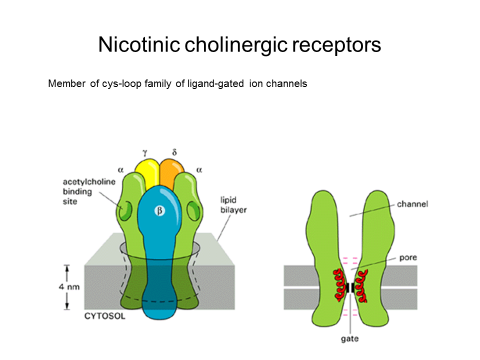
- The nicotinic ACh receptor is a fast acting molecular switch
- An oligomeric protein composed of
- an extracellular ligand binding domain
- a gated, membrane spanning pore
- The gate is opened by the binding of neurotransmitter released from the nerve terminal triggering a transient conformational change in the membrane spanning pore
- Ions flow selectively through the pore leading to a change in membrane potential
Muscarinic receptors
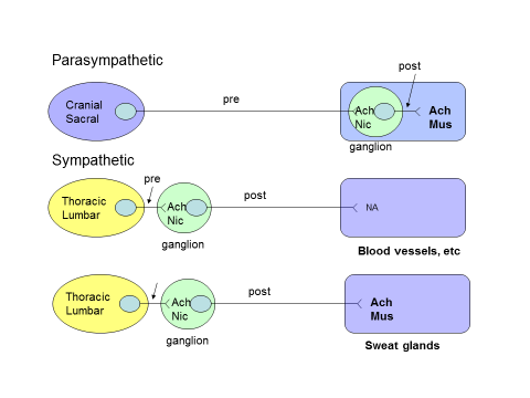
- Muscarinic receptors are located postsynaptically at the parasympathetic neuroeffector junction.
- Muscarinic receptors can either increase or decrease the activity of the effector cells.
- Muscarinic receptors are also located postsynaptically at the neuroeffector junction of sympathetic fibres in sweat glands, where they increase sweating.
- Another important site of muscarinic receptors is on endothelial cells of blood vessels. Although these muscarinic receptors are not innervated by cholinergic nerve fibres, they are sensitive to circulating molecules.
5 types of muscarinic receptors have been identified (M1-M5)
- All are G-protein coupled receptors
- Ligand binding induces a conformational change that causes the receptor to interact with a specific G-protein
- M1, M3 and M5 receptors are coupled to the activation of phospholipase C (producing inositol triphosphate and DAG as the second messengers)
- M2 and M4 subtypes are negatively coupled to adenylyl cyclase leading to reduction of cAMP formation
Clinical uses of muscarinic antagonists
- Treatment of sinus bradycardia: Intravenous atropine
- To dilate the pupil: Tropicamide eye drops
- Prevent motion sickness: Hyoscine, oral or transdermal
- Asthma: Ipratropium by inhalation
- Relaxation of GI smooth muscle: Mebeverine, oral
Noradrenergic Receptors
Noradrenergic transmission
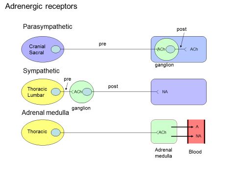
- Noradrenaline and adrenaline are catcholamines synthesised from tyrosine
- Sympathetic nerve terminals account for all of the noradrenaline in peripheral tissues (except adrenal gland)
- Heart, spleen, vas deferens and some blood vessels are rich in NA (5-50 nmol/g tissue)
- NA is stored in vesicles at the nerve terminals
- Depolarisation of the nerve terminal opens calcium channels and the entry of Ca2+ promotes fusion and discharge of synaptic vesicles
- NA release is affected by a variety of substances acting on presynaptic receptors, including autoinhibitory feedback by NA
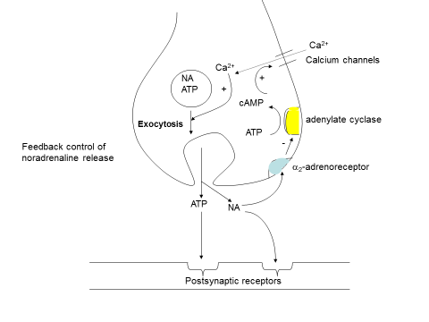
Classification of adrenergic receptors
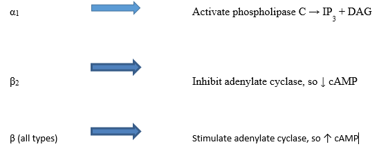
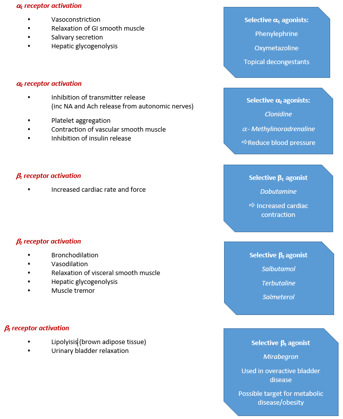
2 types- α and ß
Originally classified on strength of response to different catecholamines
- α Noradrenaline > adrenaline > isoprenaline
- ß Isoprenaline > adrenaline > noradrenaline
All adrenoreceptors are G-Protein coupled receptors
Copyright eBook 2019, University of Leeds, Leeds Institute of Medical Education.