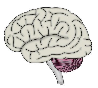
The central nervous system consists of the brain and the spinal cord. The brain is protected within the skull, and the spinal cord is protected within the vertebrae. Both the brain and spinal cord are enclosed within the meninges.
The brain is divided into the following regions:
The ventricles are the spaces in the brain and the spinal cord where cerebrospinal fluid is produced and circulated.
The cerebral hemispheres form the largest part of the brain, and can be further subdivided into four lobes; frontal, parietal, occipital and temporal. The anterior cerebral artery, middle cerebral artery and posterior cerebral artery supply the cerebral hemispheres. Obstruction or occlusion of blood flow through a cerebral artery causes ischaemic damage to the cerebral tissue that the artery supplies. Clinically this manifests as an ‘ischaemic’ stroke resulting in a focal neurological deficit.
Raised intracranial pressure can occur in children of all ages. In babies before the closure of the sutures a significantly increased intracranial volume can be reached without increasing the intracranial pressure (due to expansion of the sutures). However in children whose sutures are fused there is no room for expansion and if an increase in the intracranial volume occurs due to swelling or blockage to the normal drainage of CSF, an increase in intracranial pressure (ICP) ensues. This increased pressure will effectively “squeeze” the brain and can result in cerebellar tonsil herniation (also known as coning) where the brain is forced down through the foramen magnum. Persistent systemic hypertension is a reliable sign of raised ICP in children. Hypertension is otherwise quite uncommon in children.
A coma can be defined as a state in which the patient is totally unaware of themselves or the external surroundings. The patient is unable to respond to external stimulation. A coma usually results from global impairment of the cerebral hemispheres and/or the reticular activating system.